
Access to all articles, new health classes, discounts in our store, and more!
The Responsibility of the Management for the Effect of Focal Infection (Especially Dental) on the Life, Health, and Efficiency of the Employee
Read before the National Safety Council, Cleveland, Ohio, September 1925. Published in Dental Items of Interest, December 1925.
* * *
A new truth is a new sense, for with it an individual can see things which he could not see before receiving that new truth and which others cannot see who have not learned that new truth. But new truths come only to prepared minds. The purpose of this association, in its ultimate analysis, is to correlate and present new truths, which, when they become new senses, will conserve human life.
You have asked me to discuss the relation of focal infections, especially dental, to this matter of health conservation, and the responsibility of the management of an institution, or corporation, with regard to their associated assistants, and employees. The fundamental principle, that I am my brother’s keeper, compels me to realize that when I have a great new truth, which affects the well-being of my fellow man, it is not only my privilege but my duty to bring this information to his attention, and this fundamental principle of social progress and moral sense relates just as much to the president, the superintendent, and the foreman of a group of men and women, as it does to me as a research worker, and this is the direct problem which I am to discuss with you today.
In this communication I shall discuss this problem from the viewpoint of the opportunities and responsibilities of those persons who have the relationship to groups of working people of benefactors, while serving in their capacities as directors and supervisors. However, while I shall stress the service feature, I am glad to state my confidence that as a financial investment it seems to have been demonstrated to be not only a logical, but a very economical procedure, for no salvage can have greater value than the salvage of trained experts who frequently have required years for development.
While it is important as a money-saving proposition to prevent dental caries and dental abscess, in order to make unnecessary the loss of time and lowered efficiency of the individual during the period of his suffering, it is infinitely more important to anticipate and prevent those degenerations of fundamental organs and tissues, which are hastened in their development and frequently largely caused by the injuries to the defensive forces of the body, particularly the blood stream, and which often exist for months or years as a distinct variance from normal of the chemical constituents of the blood, apparently largely as the result of the continued presence in the body of chronic focal infections, such as dental infections.
In the light of the newer knowledge, the procedure indicated involves, not only a physical examination, but likewise a chemical analysis of the blood in order to detect these serious disturbances (serious because they exist over an extended period) and by this means not only secure evidence of the probable presence of chronic focal infections, but suggestions for reinforcing the patient at the point at which he is likely to break down later. Chronic focal infections not only produce microorganisms to invade other tissues, but toxic material which directly disturbs the balance of various factors of the blood, particularly the calcium and its compounds, which changes directly reduce the functioning, not only of organs and tissues in normal metabolism, but particularly of defensive mechanisms of the blood, as will be illustrated presently.
The health program of large corporations will therefore include a very careful study of the evidences of impending break, as they appear in the blood stream, before the symptoms of nervous breakdown, heart, kidney, eye, and many other disfunctions, which may be in the process of development and aggravation, partly or largely as the result of chronic focal infections, particularly dental focal infections.
The practical application of these principles, which my researches have developed, has been made by one of the large concerns, which has installed, in accordance with these general outlines, a department of blood chemistry in conjunction with the routine physical examination, and I am anticipating that their representative will take part in this discussion and report directly to you the results which they have achieved and the progress which they are making.
Our viewpoint has changed very materially from thinking of the dangers of injury from dental focal infections as being absent in all those individuals not showing localized disturbances, such as rheumatism, heart, kidney involvement, etc., to a recognition that these may be in part, or even large part, symptoms of an over-long continued load on the mechanisms of defense, accompanied by a final break in one or more organs, or tissues of the body.
As we see it now, the changes in the blood stream antedate the changes in the various organs and tissues, and may be recognized there, long before the physical symptoms localize as heart, kidney, or joint involvement as a disturbed function. I shall accordingly present first a group of typical lesions, which may be aggravated, or largely induced, by long-continued dental infections, or even by acute localized infections of the types that we are considering, and shall follow these with typical illustrations of the changes in the blood stream.
Altered Views on Interpretation of Radiographs.
As a preface to this, let me state a radical change in viewpoint relative to the nature and significance of the local expression of a dental focal infection. The general concept and basis of practice of the medical and dental professions has been based on the presumption that the radiograph can indicate the danger by revealing the quantity of infection, on the presumption that the extent of rarefaction, or decalcification about a root end, being the result of bacterial activity around the root end, therefore constitutes a measure of the quantity of that infection.
We now believe that the organisms, in an infected tooth, do not directly cause the absorption of the alveolar bone about the apex, except as they stimulate the mechanisms of the defense of the patient, whereby the patient, because of his or her good capacity for reaction proceeds to establish a zone of immunity; in other words, establishes a quarantine station between the tooth and the patient’s body. This we call a granuloma; and with a given dental infection–such, for example, as an infected non-vital pulp–that individual, who has a high defense, produces a large chamber in the bone; whereas, that individual with a poor capacity for reaction because of a low defense, produces a much smaller chamber, or a zone of radiolucency to the x-ray pictures.
We must therefore radically modify our viewpoints, since in the light of this newer knowledge, the individuals who most needed to have dental infections removed, often escaped having it done through the mistaken belief, that since there was little radiographic evidence, there was little infection, when the patient was in greater need for the surgical interference than one in another group capable of producing very extensive decalcification as part of the mechanism of defense.
Still another important change in our viewpoint is that for the same reason that we cannot photograph a dog behind a bush, we cannot photograph a zone of rarefaction about a root end, which is behind a zone of calcification; and it follows, that those individuals with a high defense have a marked tendency continually to decalcify in the presence of an irritant; and those with a low defense have continually a marked tendency to a limited decalcification, with a definite tendency to calcification about the zone of decalcification. This latter zone of calcification tends definitely to obstruct and obscure the radiographic view of periapical rarefaction.
Classification of Patients.
In general we can divide all our patients into three groups on the basis of how they react in the presence of a given irritant, such as a definite type of dental infection. We can also classify them into three groups on the basis of whether they do, or do not, tend definitely to have rheumatic group lesions in themselves and their families.
It is very significant and important that we put the same individuals in the various three groups by either of these two methods, for those individuals, with a marked tendency to decalcify, and who have the large areas, prove to be the individuals who have the rheumatic group diseases; and, further, that those, who have the large zones of rarefaction, surrounded by a zone of calcification, prove to be those who have tended not to have the rheumatic group diseases, but under the stress of overload have had a break in their defense, and who now become susceptible to these affections of certain types; and, third, those with a very marked tendency to calcify with a limited tendency to decalcify, prove to be those who through life have had a marked tendency, not only in themselves, but in their families, to have the rheumatic group affections.
I will present a limited number of illustrations of the types of affections which seem to have direct relation to dental focal infections, but it will be clearly understood that the limited space of this article will not allow more than a few suggestions as illustrations. I have already presented this in extensive detail in a two-volume work on dental infections. (Volume I–Dental Infections, Oral and Systemic. Volume II–Dental Infections and the Degenerative Diseases. Penton Publishing Co.; Cleveland.)
A Case Record, Anemia.
If we will consider first some blood stream changes, we will think at once of the heart involvements and anemias. The latter is illustrated by the following case. A young man was brought from one of the hospitals, suffering from spontaneous hemorrhage, chiefly of the gums. He had had a hemorrhage in the internal ear about two weeks previously which caused complete deafness. The bleeding had been going on for two or three months, and on two occasions had been so severe that he became practically unconscious from loss of blood, and had blood transfusions, the beneficial effects of which, however, lasted only a few days.
When brought to me, the blood was oozing from the gums and dropping from his mouth at the rate of about one drop in two seconds, which he said was about the rate it had been for the last six weeks. He was ashen in color. Careful examination showed that he had five pulpless, root-filled teeth, which did not show extensive absorption. They were what are commonly called x-ray negative. It was disclosed that the hemorrhage was most severe around these teeth. The clotting time of his blood was ten minutes.
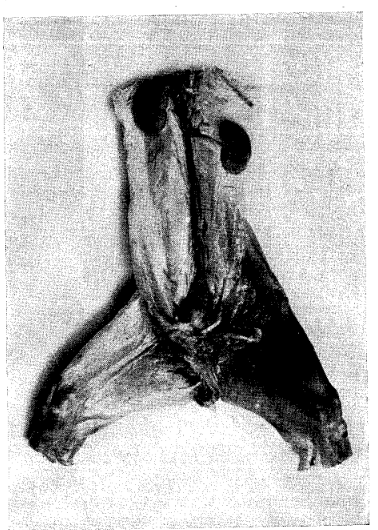
Fig. 1. Fatal, multiple, spontaneous hemorrhages in rabbit, produced by culture from tooth of patient suffering from grave oozing of blood from gums.
One of these teeth was removed, the socket packed to control the hemorrhage, and he was retained in our ward for very careful attention and observation. A culture was grown from the interior of this tooth, which was inoculated the following day into a series of rabbits, one of which is shown in Fig. 1. The rabbit died in about twenty hours from spontaneous hemorrhages all through its system, as illustrated in the thighs and kidneys. Several of the animals developed such hemorrhages and their clotting time changed from one to two minutes as normal, to six, eight, and ten minutes, due to the presence in their system of this organism. The amount of culture inoculated was 1 cc. of an eighteen hour growth.
With the removal of one after another of these pulpless teeth his clotting time changed progressively toward normal, and, in two weeks’ time was down to three minutes. The spontaneous hemorrhages had entirely ceased, and in five weeks’ time he was back at his regular employment.
In the light of our newer knowledge regarding calcium, to which we will refer later though briefly, we have an indication of the role of the particular type of infection that was involved in this case. We have had rabbits die in eight hours, from spontaneous hemorrhages from such strains, taken from the teeth of individuals suffering from hemorrhages.
We cannot cite more than one illustration of various types and space will permit only a few types.
A Case Record, Heart Involvement.
In further consideration of the circulatory system disturbances, let us consider heart involvements. A boy was brought for study, who had developed acute rheumatism. He had had a severe toothache, about six weeks previously. It was not known that he had a heart involvement, until our examination revealed a very severe endocarditis. The pulp was still vital in the tooth, and though the caries was deep, the pulp was not exposed.
On removal of the tooth, a culture was made and inoculated into thirty rabbits. Twenty-eight of the thirty (or 93%) developed heart involvement, and thirty of the thirty (or 100%) developed acute rheumatism.
The boy died about six months later of endocarditis, which affection had caused other deaths in the family. His inherited lack of defense for this type of infection, particularly as it related to heart, doomed him before birth, unless the intelligent supervision of his home and his employer could come to his rescue, with a program that would prevent his becoming a prey at the point of his inherited lowered immunity.
Quantity of Infection Which Is Dangerous.
The quantity of infection adequate to do damage is apparently very much less than we have previously been led to believe. For example, two teeth of a young man twenty-six years of age, suffering from acute endocarditis frequently recurring, were crushed, and the toxic material and organisms washed from them and injected into a rabbit. The rabbit died in a few weeks’ time from acute endocarditis, with a valvular vegetation one inch long. This heart is shown in Fig. 2. The quantity of organisms, by weight, was determined by making a count from a sample of the material injected, and found to be less than the millionth part of a gram, and yet they were sufficient, because of their extreme localization qualities, not only to attack the heart, but to produce the death of the animal with typical endocarditis.
Fig. 2. Valvular vegetation in heart of rabbit which died of endocarditis after being inoculated with washing of two crushed teeth from a patient suffering from acute endocarditis.
Passing to affections of the nervous system, it is of interest to note that in over two thousand rabbits inoculated with cultures from dental origin, paralysis of all or part of the body has not occurred in one-half of one per cent of the total inoculations with strains from the teeth of individuals not suffering from acute nervous system disturbances. However, when individuals are suffering from an acute nervous disturbance, some strains show very definite localization, as illustrated in the following case.
A Case Record, Nervousness.
A young lady presented with attacks of uncontrollable nervous irritability, recurring. An official of the firm, for whom she was working, called me on the phone and stated that he was sending her out for examination, but said he wished to offer the suggestion that she liked sympathy so much that she was having these spells for that purpose, and he wished accordingly to put me wise.
A tooth was extracted of the type which formerly we would not have thought serious, but in the light of our newer knowledge we know to be the most serious type. A culture was grown from its interior, and inoculated into four rabbits. Three of the rabbits were paralyzed from the centers of their spines backward, as shown in the illustration, Fig. 3. They dragged their hind legs as though they did not belong to them.
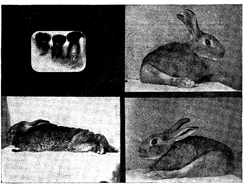
Fig. 3. Three rabbits paralyzed by intravenous injection of culture from tooth shown. Patient seriously suffering from nervous attacks.
One of these animals is shown in dissection and radiograph in Fig. 4, and it will be noted that where the infection has attacked the spinal column, the spinal cord has come to be compressed from the building on of bone in the process of condensing osteitis. Please note in the radiograph that there is no chamber of rarefaction, but on the contrary one of increased density. A, shows the paralyzed rabbit, B, a dissection of the spinal column showing the spinal cord as it is compressed, C, a photograph of the ventral surface of the spine, revealing the involved cartilage, and D the radiographic appearance of the lesion.
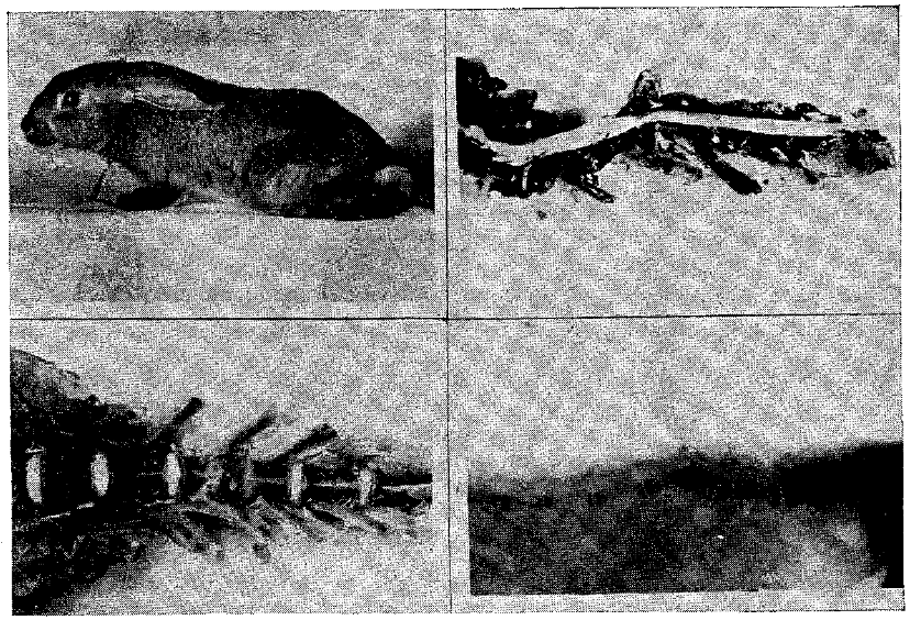
Fig. 4. Paralyzed rabbit. A. Loss of control from center of spine back. B. Lateral view of dissection, showing compression of cord. C. Ventral view, showing destroyed cartilage. D. Radiograph showing condensation, not rarefaction.
Tetanic Spasms.
Fig. 5 shows two rabbits inoculated intradurally, each with a different culture, but both from the cultures from teeth of patients. One patient showed no systemic disturbance. The other had recurring convulsive tetanic spasms of encephalitis. With her recurring attacks, she would brace herself in her chair, her arms becoming rigid, and violent spasms of her muscles of mastication produced a grinding and crunching of her teeth that could be heard in the surrounding rooms. The attacks would last about three to five minutes.
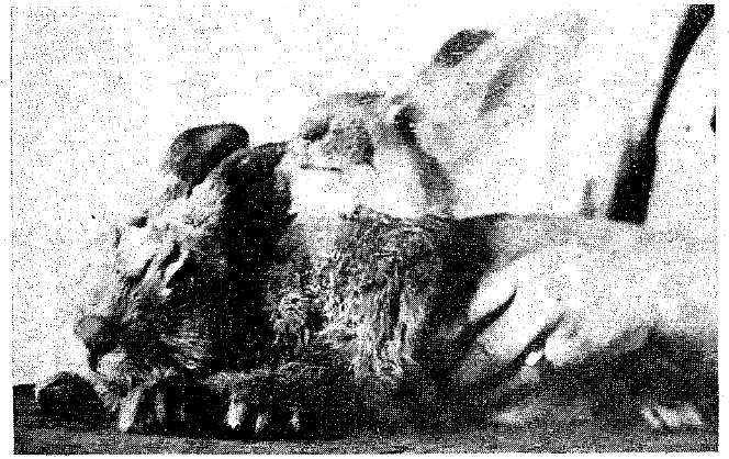
Fig. 5. Rabbit with convulsive spasms, from inoculation with culture from tooth of patient with encephalitis and suffering similarly.
The culture from her tooth inoculated into one of these rabbits produced, as revealed in the photograph, similar tetanic spasms, for the rabbit is seen in a convulsion with its four feet rigid, its head thrown back, and its muscles of mastication spasmodically contracting, with so violent a grinding noise that it could be heard many feet from the animal. The spasms similarly lasted about three to five minutes. The animal died in one of them within five hours after the inoculation.
Torticollis
Among the many lesions, directly and indirectly related to dental infections, are those of the cervical nerves of the muscles of the neck. These may produce typical torticollis, or may only produce discomfort in the patient. Usually the patients think that they have caught cold in the neck, the facts being that a draft striking the tissues lowered the local resistance so that the tissues readily became a prey to the toxic and bacterial invasion.
Such a case is illustrated in Fig. 6, which shows four rabbits, three with their heads drawn back, one with a very extensive myositis of the hips. The patient from whom the cultures were taken had been suffering for eight days with an acute torticollis, which had been recurring in mild form for two years, but with increasing severity. The infected roots of a tooth were extracted, and a culture inoculated into a group of rabbits, many of which developed acute nervous affections with displacement of the head. The rabbit in the upper left was in this group.
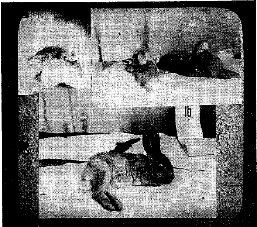
Fig. 6. The two rabbits with heads drawn back were inoculated, one from culture from the patient’s tooth, and the other from a culture from the neck muscle of the patient suffering severely from stiff neck. The lower rabbit is paralyzed in its hips.
A piece of the patient’s neck muscle was taken under a local anesthetic and sectioned, and showed streptococci in the musculature. A piece of the muscle was ground up with sand and cultured, and the culture, which proved to be Streptococcus, was inoculated into a second group of rabbits, several of which developed similar symptoms to those inoculated with the tooth culture. One of these is shown the second from the left in the upper part of Fig. 6.
The removal of these teeth completely relieved this patient of her disturbance for a period of two years, when the symptoms developed again but on the opposite side of her head. Investigation showed an involvement of the pulp of a tooth from deep caries underneath the crown of a bridge. Within five hours after the removal of this tooth the symptoms had disappeared.
Another striking illustration of this elective localization quality will be seen in Fig. 7, in which four views of a rabbit are shown, which had been inoculated with the culture from a tooth of a patient suffering from a recurring involvement of the muscles of the side of her head, neck, and shoulder, but which entirely disappeared after the removal of the dental infection. This rabbit’s head was continually drawn to one side, but placing the rabbit under an anesthetic, releasing the nerve control, allowed the head to become entirely flexible and relaxed only to return again immediately on the recovery from the anesthetic.
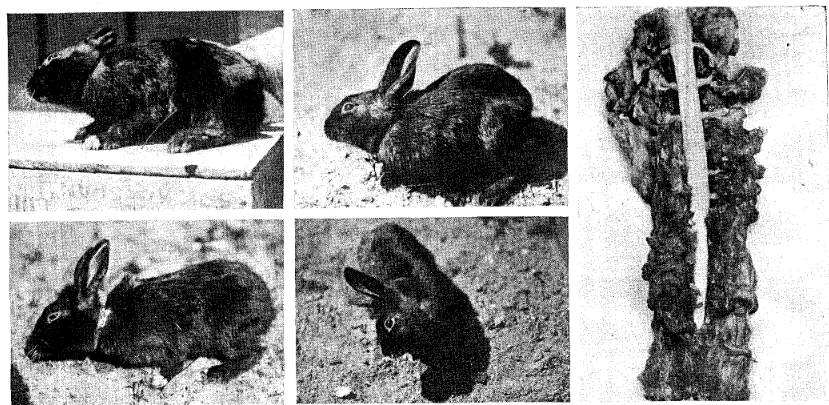
Fig. 7. Four views of a rabbit with wry neck from inoculation with dental culture from a patient suffering from wry neck.
Nephritis.
We have come to look upon kidney irritations, including some of the types of nephritis or Bright’s disease, as having direct connection with dental infection. Fig. 8 shows the greatly enlarged kidneys of a rabbit; five times their normal size, as shown in comparison with a normal rabbit kidney seen beside the abnormal one. This rabbit was inoculated with the cultures from the teeth of a young woman who had been almost completely incapacitated by Bright’s disease, but who greatly needed to work, her husband being dead and she having a child to support. Within a few weeks after the removal of these dental infections, the albumen had entirely disappeared from her urine, and her nephritis was so completely relieved that for six years she has been able to work continuously, full time. This illustration shows sections of the kidney. Within two weeks the rabbit had casts in its urine, and at the time of posting just before its death the kidneys had developed the extreme parenchymatous nephritis shown in the illustration.
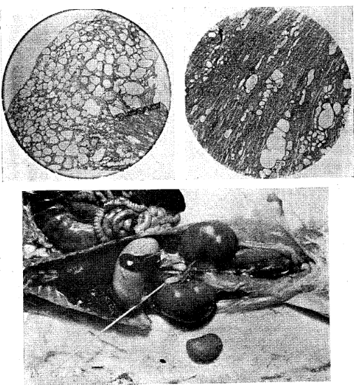
Fig. 8. Diseased kidneys of a rabbit with nephritis five times normal size. (See a normal below for comparison.) Upper are sections of same.
Arthritis.
Of the affections produced or aggravated by dental infections, the arthritides are probably among the most frequent. We now recognize two principal types, one of which is quite frequently related to focal infection. Fig. 9 shows a boy of sixteen, seriously affected, for he had been suffering acutely for four years, most of which time he had been bedridden and practically helpless. His jaw was ankylosed, so that he could only take liquids and his pain was so great that he cried daily by the hour.
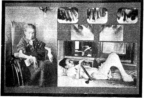
Fig. 9. This very severe arthritis was getting progressively worse. It progressively improved after the removal of dental infections.
With the removal of his dental infections he so greatly improved that he not only was free from his acute involvements, and could open his jaws quite naturally, but was able to work with his hands. Fig. 10 shows the same boy beside an aeroplane and some fancy carvings that he has made with the same hands, which before were so crippled.

Fig. 10. Handiwork made with these formerly nearly useless hands.
A most important and enlightening aspect of this case, and which is typical of this type of patient, is the nature of the dental pathology, as revealed in the radiograph. Fig. 11 shows an upper lateral incisor (the center of the three in the picture) which had a putrescent pulp, but which with all its infection showed very little radiographic evidence of periapical disturbance.
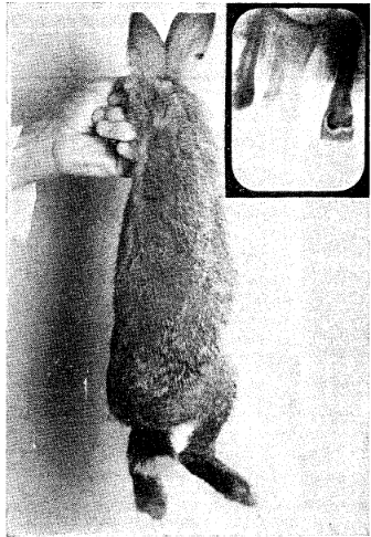
Fig. 11. A tooth from boy in previous illustration. Note absence of apical rarefaction. This rabbit has acute rheumatism from culture from this tooth.
The culture taken from this tooth, however, inoculated into the rabbit shown, produced acute rheumatism in twenty-four hours, which in forty-eight hours induced the animal to go on three legs. The hind leg, shown raised, has acute arthritic infection in the joints.
Affections of the Eyes.
Of the many affections which we should present, most of which we cannot for lack of time, will be those of the eyes. Figure 12 shows different views of the eyes of a rabbit inoculated with a culture from the teeth of a patient suffering with acute eye involvement affecting only one eye. The dental conditions were not such as would be considered even suspicious, under the old standards of diagnosis, for they showed no periapical involvement. It is most remarkable, however, that four of the five rabbits inoculated with this culture developed acute involvement of one, or both eyes. The normal eye of this rabbit is shown to the right and the diseased to the left. Two stages of the development of acute corneal ulcer, which resulted in the complete blindness of the eye, are shown.
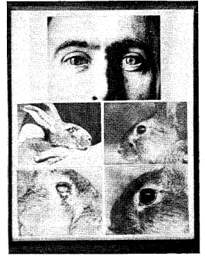
Fig. 12. One eye of patient had acute retinitis. Four of five rabbits inoculated with dental culture developed acute eye involvements; one was blind. Eyes to right are normal; to left, affected.
Fig. 13 shows the pus running out of a rabbit’s eye forty-eight hours after it was inoculated with a culture from the tooth of a patient who was rapidly losing the vision of the eye, and which vision had greatly improved in ten days’ time and returned practically to normal, after the removal of the dental infection, from which teeth the culture was taken for inoculation into this animal. Space will not permit the presentation of further typical cases.
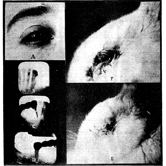
Fig. 13. Extreme eye involvement of Case No. 861. B, C, and D. Dental conditions. E and F. Progressive stages of acute involvement in rabbit’s eye, inoculated with culture from these teeth.
Factors of Immunity and Susceptibility
In a part of the limited remaining time I wish to discuss some of the fundamental factors of immunity and susceptibility to injury from dental infections. When an extracted infected tooth is placed beneath the skin of a rabbit, it has a very different effect from the placing of a sterile tooth. The latter is easily encapsulated and usually produces very little local or systemic disturbance. An infected tooth, however, slowly breaks down the defense of the animal, with a series of blood changes which are quite constant and characteristic.
Similar results are obtained frequently by injecting into the circulation the washings of a crushed tooth, and in some instances the injection of the bacteria-free filtered washings of an infected tooth will produce the typical conditions. Fig. 14 shows two rabbits, brothers, one of which was inoculated with the washings from an infected tooth and the other was not. The control gained fifteen per cent in five weeks’ time, during which time the one that was inoculated, which is shown above, lost thirty-five per cent in weight and died with typical marasmus and blood stream changes.
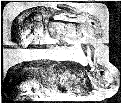
Fig. 14. Upper rabbit had been inoculated with washing from crushed tooth. It lost 35 per cent in five weeks, while its normal brother gained 15 per cent.
We have made several hundred rabbit studies of this type, and, in approximately seventy-five per cent of the cases the animals have died within a few days or weeks. There is a characteristic depression of the polymorphonuclears with a corresponding increase of the percentage of small lymphocytes. This depression of polymorphonuclears and increase of lymphocytes amounts on an average to about seventeen per cent.
Consideration of the Active Calcium.
Accompanying this disturbance of balance of the blood cells and a primary factor in its causation is a depression of the active calcium. This is shown in Fig. 15, in which a series of animals are included. Accompanying a more or less extreme loss in weight there has been a depression of the active calcium, ranging from 2 to 6 mgs., or from 10 to nearly 50 per cent. This change in calcium is probably one of the most important of the effects of chronic focal infections, for cellular activity of the various organs and tissues of the body is dependent upon a quite exactly balanced ratio between the various chemicals involved, and also it requires a relatively constant level for each of these, particularly of the calcium.
Depression of Ionic Calcium by Implanting Infected Teeth
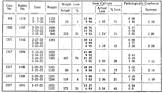
Fig. 15. Showing depression of blood calcium in rabbits with an infected tooth planted beneath the skin.
In Fig. 16 will be seen a typical example in a single rabbit of these blood changes. In this case, a dental culture was inoculated. In six days the polymorphonuclears decreased from 57 per cent to 31; the small lymphocytes increased from 34 to 62 per cent, a change of approximately 26 with each. While this was taking place, the active or ionic calcium decreased from 11.5 to 8.05. When the active calcium is depressed to 40 or 50 per cent of its normal, the animals tend to go into convulsions and usually die in a short time. For those who are interested in calcium determinations, it will be of interest to note that while the total calcium increased from 13 to 13.06, the active, or ionic, decreased from 11.5 to 9.4, demonstrating, as we have now so abundantly done, that simply the determining of the total calcium is very inadequate for getting the true picture.
Comparison of Changes in Ionic Calcium and Blood Morphology, due to Culture Inoculations
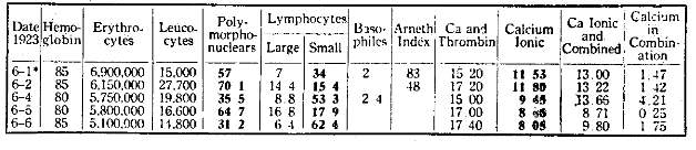
Fig. 16. Depression of a rabbit’s polymorphonuclear blood cells and increase of lymphocytes and also of blood calcium, from inoculation with dental culture.
A more detailed and graphic expression of the effect of tooth implantation is revealed in the following case. Rabbit No. 1356 had a tooth implanted beneath its skin and various of the calcium factors were determined. In nine days’ time it was found that the total calcium decreased from 13.3 to 12.2. During this time the active calcium decreased from 10.5 to 6.1. The diffusible calcium decreased from 4.9 to 4.7; the non diffusible calcium from 8.4 to 7.4; the inorganic phosphorus from 4.5 to 2.9. The effect of this infected tooth beneath the skin of this rabbit is typical of many determinations that we have made of this same problem. The animal may or may not become seriously languid sometime before a crisis comes. Frequently, when the active calcium goes down to around 5 or 6 mgs., the animals go into convulsions and die even though the stomach is well filled with recently eaten food.
When chronic dental infection exists in the body for an extended period, it produces changes in the levels of various chemicals of the blood, which may be accompanied by little or no localized tissue change anywhere in the body. The individual has a sense of languidness. It is as though the brakes were on. They are usually underweight. They do not know it, but they are often heading toward a break, which may come in the nervous system, or in some of the organs, or in the skeleton and musculature. Usually the condition exists in the blood stream for a few or many months before the serious break comes. If that break shall have induced the destruction of an organ or tissue that is fundamental to life, such as the heart or kidney, it produces a grave situation. If it be in the eye, it may mean the partial or complete, temporary or permanent, loss of vision; if in the nervous system, the entire disposition may be changed. As we now understand these various affections, which we know as degenerative diseases, they are in part, and often large part, expressions and therefore symptoms and end effects of the primary changes in the blood stream. The unfortunate possessor of a heart valve that has been once involved, tends, as is common knowledge, to have a recurrence, and such an individual is usually handicapped, more or less seriously, for life and usually has life shortened. In the light of this newer knowledge the better treatment of the heart affection will be its prevention.
Libman and others have shown that the organism involved in subacute endocarditis is in 95 per cent of the cases Streptococcus viridans. Our work has shown that in more than 99 per cent of cases, the organism growing in infected teeth is the Streptococcus viridans, though we now subdivide it into several sub-groupings.
Time has not permitted an analysis of the role of contributing factors as overloads, such as influenza, poor nourishment, exposure, pregnancy, heredity, etc. I have discussed these at length in the two volumes above mentioned and in various papers.1 In these communications I have shown that a considerable percentage of humanity is born with a lower than normal capacity for combatting streptococcal infection, which is the reason, for example, why we have recognized that heart disease runs in various families, and similarly with other affections. Such individuals, as are so handicapped, and it probably constitutes 20 to 25 per cent of the total population in a community, not only have a greater susceptibility to dental caries, and therefore to dental focal infections, but require an entirely different type of protection, if they are to live a life of normal comfort, duration, and usefulness. They do not and cannot have the knowledge that will enable them to carry out such a program. It therefore becomes a matter of necessity that others shall furnish them this care. We have accordingly, then, the prime responsibility as keepers of our brothers, that we shall provide means for the protection of those, who by inheritance and exposure are so handicapped.
A group of such children, all having chorea (Saint Vitus’ dance) were taken from London to a seashore vacation. They all recovered from their chorea. They were then taken back to their home environment. In six months’ time they were all examined again and over 60 per cent had developed heart involvements, which they did not have when they were taken to the seashore.
It is a physical impossibility for such individuals to grow up in the handicaps that modern home and working conditions provide without a premature break. Just as the Eskimos do not die of measles unless they are exposed to measles, though practically all Eskimos exposed to measles die, just so individuals without a normal defense against streptococcal infections may live a relatively normal life if they do not have a source for that infection, but are endangered, if not doomed, if they should develop such; and whether they break or not depends upon one other factor–namely, the extent of the overloads–which may be unsatisfactory working conditions.
I have had, for example, in my care a woman with multiple deforming arthritis so advanced that she cannot move a hand or foot nor scarcely any joint of her body, whose acute first attack came on at the time that she was sorting harness castings in a room improperly warmed, and these were brought in chilled from another and unwarm part of the building. At the time she had dental infections, which several factors seem in the light of our newer knowledge to have created, with her inherited susceptibility, all that was necessary to bring about this tragic situation that has made her a bedridden invalid for now approximately twenty years.
Can we say of this generation that the management was not responsible because they did not know? One in ten of approximately all the funerals of the civilized countries of the world is a death due to heart involvement. We have a conscience and a program for prevention of tuberculosis, but we do not have such for the prevention of heart involvements. In one of our modern communities, the diseases of adults which unnecessarily shortened life during 1923 were of a total of 470, diseases of the heart 112, nearly 25 per cent, cerebral hemorrhage (apoplexy) 57, cancer 56, pneumonia and influenza 44, diseases of the arteries 26, diseases of kidneys 22, diabetes 16, tuberculosis, all forms, only 10, all other causes 127.
Is it not a sad incident to record for this generation that we have not awakened up to the fact that the degenerative diseases of the heart are largely preventable? While you, who have the responsibility of the people in your care, have a glorious opportunity for saving money for your corporation by conserving working efficiency through the prevention of focal infections, it is my personal belief that presently, if not already, you shall be held in large part responsible for not only the physical efficiency but for the morbidity and tenure of life as well.
References Cited:
- “Dental Infections and Related Degenerative Diseases, Some Structural and Biochemical Factors.” J. A. M. A., Jan. 24, 1925, Vol. 84, pp. 254-259. “Fundamentals Suggested by Recent Researches for Diagnosis, Prognosis, and Treatment of Dental Focal Infections.” J. A. D. A., June, 1925.