
Access to all articles, new health classes, discounts in our store, and more!
Iritis Produced in Rabbits’ Eyes by the Intravenous Injection of Crude and Purified Cultures of Bacteria Isolated from Patients with Certain Inflammatory Eye Diseases
Read before the Association for Research in Ophthalmology, Kansas City, Missouri, May 12, 1936. Published in American Journal of Ophthalmology, Vol. 19, No. 12, December 1936. Co-Authors: Conrad Berens, M.D., and Edith L. Nilson.
* * *
Iritis was produced in rabbits by the intravenous injection of either primary or purified cultures from 19 to 21 patients with acute or chronic eye diseases, and in 11 of 14 controls (laboratory assistants, healthy children, and patients with arthritis and thyrotoxicosis).
Positive results were obtained with various microorganisms as follows: streptococci (alpha, beta, and gamma types), staphylococci (albus and aureus), colon bacilli, non-lactose fermenters, enterococci, and Friedlander bacilli.
Iritis was produced by 44 percent of 61 purified strains of streptococci from patients with eye disease as compared with 29 percent of 69 strains from persons in the control group.
Of the total of 134 cultures from patients with eye disease, 36 percent produced iritis while 17.9 percent were undetermined. Of the total of 118 cultures from persons in the control group, 29.2 percent produced iritis in rabbits, while 21.5 percent were undetermined. (From the Lighthouse Eye Clinic of the New York Association for the Blind, and the Clinical Research Laboratory. Aided by grants from the Ophthalmological Foundation, Inc.)
Because of the possible importance of the relation of focal infection to the etiology of many acute and chronic eye diseases, a knowledge of the relationship of microorganisms to the production of ocular lesions is of vital importance. The impracticability of inoculating human volunteers has made it necessary to study this problem by means of animal experimentation. Rabbits are susceptible to the pathogenic action of many bacterial species and are less expensive than primates. Therefore, they have been used extensively, even though positive findings cannot be considered conclusive evidence of a parallel relationship to ocular disease in man.
In 1932, Rosenow and Nickel1 summarized a series of experiments previously published by them and their associates on the elective localizing power in rabbits of freshly isolated streptococci and pneumococci derived from foci of infection of patients with various diseases. (The lesions produced in their earlier experiments had occurred only after several passages through animals of “laboratory” strains of these organisms.) They also reported a new series of experiments, following a somewhat similar method, in which iritis was produced by direct inoculation of primary cultures from patients suffering from acute, chronic, primary, or recurring attacks of iritis, uveitis, or iridocyclitis.
This report and the results of other investigations, such as those of Maestro,2 Zanettin,3,4 Blanc and Martin,5 Cusumano,6 Wherry and King,7 de Andrade,8 von Herrenschwand,9 Brown,10,11 Irons, Brown and Nadler,12 Meisser and Gardner,13 and Haden14 stimulated the experiments to be described in this paper.
Experimental Procedure
A series of patients with acute and chronic inflammatory eye diseases was studied bacteriologically. Because previous experiments had indicated that the nose and throat, even though symptomless,15 were the foci most frequently involved in chronic or acute diseases,16 they were chosen as the most favorable areas from which to obtain cultures. Cultures from teeth and tonsils were also used in certain instances. In most of the early experiments, separate cultures were made from the left and right nostrils but, as only minor differences were noted, subsequent cultures from both nostrils were combined.
Dextrose brain broth, made by adding approximately 3 gm. of calves’ brains to about 10 c.c. of Bacto brain-heart infusion, was used for primary cultures. Each swab was placed in a tube of this medium and incubated for 18 to 24 hours. The swab was then discarded and a loopful of the culture was spread on two blood-agar plates by means of a glass spreader.17 Following Rosenow’s suggestion, blood-agar cultures were grown anaerobically. Ordinarily there was a lighter growth on the second plate, which made it easier to find discrete colonies and to differentiate the various types. Since most of the cultures proved to be mixed growths, this was important. The primary cultures were then injected intravenously into rabbits. The organisms isolated from the blood-agar plates were purified, grown for 18 hours in brain-heart infusion and tested for toxicity* by the in-vitro methods of Chapman, Berens, and their associates.18,19,20,21 The purified cultures were then injected intravenously into albino rabbits weighing between 1,400 and 1,600 gm.
In recording the occurrence of iritis, the designations two plus (++), three plus (+++) and four plus (++++) indicate the degree of iritis produced. Two plus (++) indicates definite congestion with marked engorgement of the vessels. Three plus (+++) indicates marked congestion of the iris, marked circumcorneal congestion, edema, and clouding of the iris with or without small hemorrhages. Four plus (++++) iritis indicates the same as three plus (+++) with the addition of exudate in the anterior chamber.
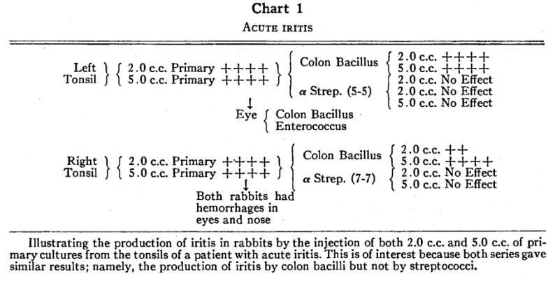
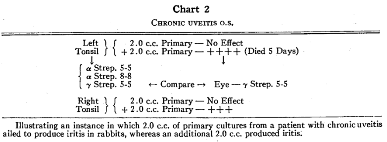
Both 2.0 c.c. and 5.0 c.c. of the primary cultures from the first cases studied produced iritis in rabbits (chart 1). For the next few cases only 2.0 c.c. of the primary cultures was used. The results were negative, even though the patients from whom the cultures were obtained had pronounced ocular symptoms. An additional 2.0 c.c. or 3.0 c.c. of the same primary cultures was therefore injected into the same rabbits within 24 hours. Positive results were obtained in a number of instances (chart 2).
As a result of these findings, the initial dose was increased to 5.0 c.c. The increased dose produced satisfactory results with throat cultures but death occurred rapidly in the majority of rabbits injected with nasal cultures. Further study led to the belief that death was due to the colon bacilli and toxic staphylococci often recovered from the nasal membranes, and to the fact that these organisms grew more luxuriantly than streptococci, which usually predominate in the throat. It was then decided to use as an initial dose 5.0 c.c. for throat cultures and 3.0 c.c. for nasal cultures, although the optimum dose for each case varies and cannot be predetermined. In the case of nasal cultures, when an injection of 3.0 c.c. did not result in death or ocular disturbance within 12 to 24 hours, an additional 2.0 c.c. or 3.0 c.c. was usually given.
The rabbits were observed at intervals commencing six hours after inoculation. Detailed examination was made after 12 to 15 hours and, if no ocular lesions were noted, again after 24 to 48 hours. The animals were then discarded. Recent observations show that iritis may appear as early as one to three hours after inoculation and subside within a few hours. In other instances, a definite iritis may not appear until the end of 10 to 12 hours. This indicates the necessity of early and more frequent observation.
When a primary culture produced a pathologic effect in the rabbit’s eyes, all the purified brain-heart-infusion cultures of the isolated organisms were inoculated into rabbits to determine, if possible, which strain or strains produced the original eye lesion. This was also done when the rabbits died too early for the appearance of eye symptoms or when they died during the night. Ordinarily, if the primary cultures showed negative results and the rabbits survived 48 hours, no other rabbits were inoculated. Conjunctivitis was ignored except when it was marked. With one exception, whenever a primary culture produced iritis in rabbits, one or more of the purified strains also produced iritis in rabbits. Iritis was produced with pure cultures of streptococci, enterococci, non-lactose fermenters (degraded colon bacilli?), colon bacilli, and staphylococci.
Chart 3 illustrates a case of hemorrhagic retinitis in which the primary cultures did not reveal significant information, but in which four of the five strains of streptococci isolated from the throat culture produced iritis in rabbits. This demonstrates the value of testing individual strains.
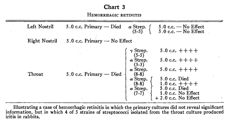
Chart 4 illustrates the findings in a case of sclerokeratitis, possibly tuberculous, in which the primary cultures killed rabbits overnight but all the purified strains of streptococci produced iritis in rabbits.
Chart 5 illustrates the production of iritis in rabbits by the intravenous injection of a pure culture of enterococcus obtained from the left nostril of a patient with recurrent uveitis.
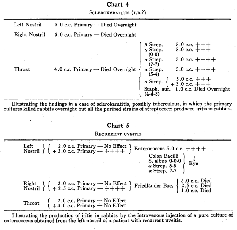
Chart 6 illustrates a case of recurrent iritis and episcleritis in which iritis was produced in rabbits by a non-lactose fermenter (possibly a degenerate strain of colon bacillus) isolated from the right nostril.
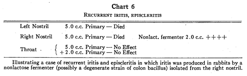
Chart 1 illustrates a case of acute iritis in which alpha streptococci and colon bacilli were isolated from the primary tonsil cultures. The colon bacilli produced iritis in rabbits while the streptococci failed to produce iritis.
Chart 7 illustrates a case of suspected chronic tuberculosis of the choroid in which Staphylococcus aureus from the left and right nostrils produced iritis in rabbits with primary cultures but failed to do so after subculture.
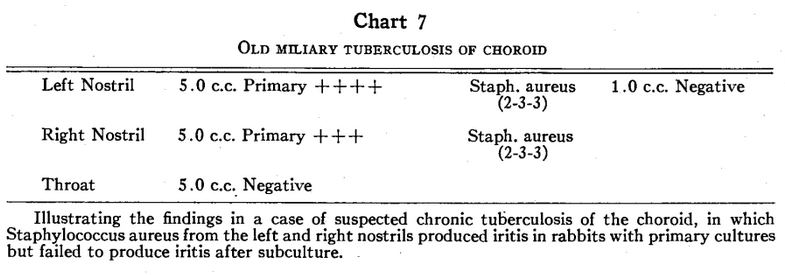
In the earlier experimental work, staphylococci produced eye disease only with the primary cultures, as shown in chart 7. Apparently the power to produce iritis was often lost before the subculture could be injected because, when it did not kill the rabbits, the eyes remained normal. Therefore, the inoculation of purified strains of staphylococci was discontinued temporarily. On resuming the testing of purified strains, iritis was produced in several instances when only 1.0 c.c. of the culture was used.
Control Experiments
Control cultures were obtained from apparently healthy persons having no obvious ocular infection. Five series of cultures were from laboratory assistants and three were from children. The remainder were from patients with chronic diseases such as arthritis, thyrotoxicosis, and so on, but with no eye disease. The findings were similar to those in patients with inflammatory eye diseases, iritis being produced by cultures of streptococci, Friedlander bacilli, staphylococci and colon bacilli, although the frequency of positive results was not quite so high.
Chart 8 illustrates a control case (laboratory assistant) in which the primary throat culture and two of the five strains of streptococci isolated from it produced iritis in rabbits.
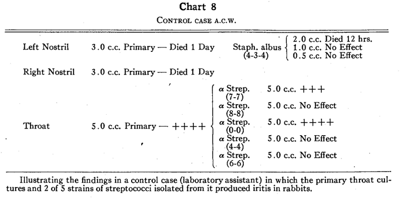
Chart 9 illustrates a control case (laboratory assistant) in which iritis was produced in rabbits by a strain of Friedlander bacillus isolated from the left nasal culture, by a strain of staphylococcus isolated from the right nasal culture, and by a strain of streptococcus isolated, from the throat culture.
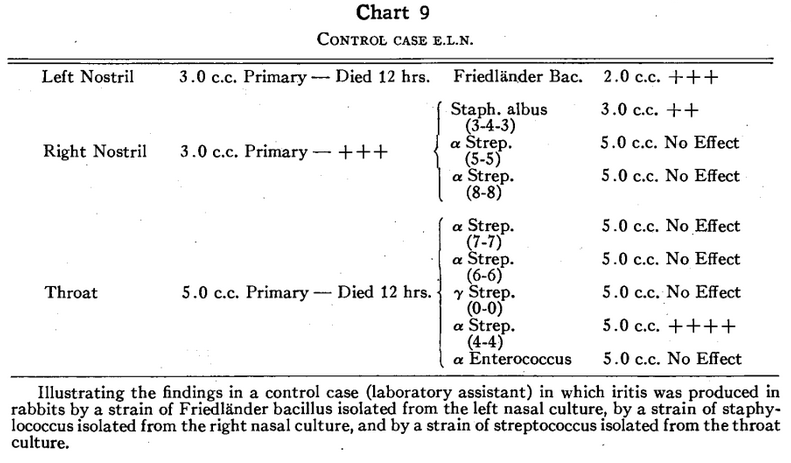
Chart 10 illustrates a control case (laboratory assistant) in which a strain of colon bacillus isolated from the primary right nasal culture produced iritis in a rabbit. This is an instance in which Staphylococcus albus produced iritis in primary culture but not in subculture.
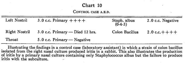
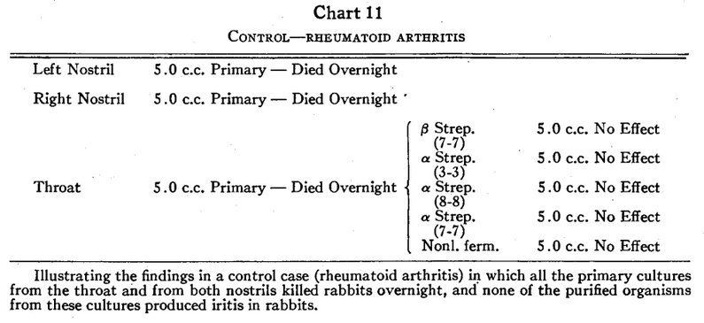
Chart 11 illustrates a control case (rheumatoid arthritis) in which all the primary cultures from the throat and from the left and right nostrils killed rabbits overnight but in which none of the organisms isolated from these cultures produced iritis in rabbits.
Method of Culturing Eyes
In the early experiments, cultures of the eyes were made in nine of the animals in which eye lesions had been produced. Two cultures were overgrown by “spreaders,” six yielded a number of different organisms, predominantly enterococci and colon bacilli, and only one yielded an organism similar to that injected intravenously. The method of obtaining the cultures may have been at fault. The eye was enucleated as soon as possible after death, placed in 50-percent alcohol for 15 minutes and drained. It was then dropped into brain-heart infusion and cut. By the following method, which is now employed, no “spreaders” have appeared on the plates. The animal is anesthetized while the inflammation is at its height, the conjunctiva is irrigated with 1:200 Metaphen solution, a 27-gauge needle attached to a tuberculin syringe is plunged through the corneoscleral margin into the aqueous and all the fluid is aspirated. The possibility of contamination is thus reduced and, because the animal is still alive, the likelihood of obtaining live pathogenic organisms free from postmortem invaders is increased. Four eyes have been cultured by this latter method and two organisms similar to those injected intravenously (streptococcus and Staphylococcus aureus) have been recovered. When an organism similar to that injected intravenously was recovered from the aqueous of the rabbit’s eyes, the organism isolated from the eye produced iritis in other rabbits. When organisms different from those injected intravenously were recovered from the aqueous they did not produce iritis in other rabbits. Eye cultures from six normal rabbits were negative.
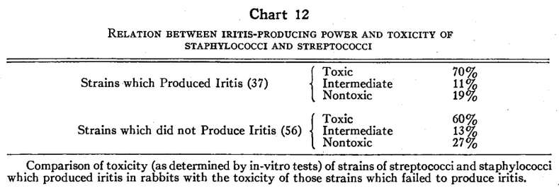
Seventy percent of the iritis-producing strains gave positive toxicity tests by the in-vitro methods, while 60 percent of the strains which did not produce iritis also gave positive in-vitro toxicity tests. Thus, the proportion of toxic strains (as judged by in-vitro tests) among those which produced iritis and those which did not produce iritis was similar (chart 12).
Discussion and Comparison of our Experimental Results with Those of Other Investigators
The subject of the production of iritis in rabbits by various microorganisms is complex. It is further complicated by the use of various methods by different investigators. For example, Zanettin3 and Brown22 endeavored to enhance the iritis-producing power of organisms by growing them in association with uveal tissue but reached opposite conclusions. Maestro2 tried to produce oculotropic properties in streptococci by passage through normal rabbits’ eyes. Cusumanos sought this effect by numerous passages of Streptococcus viridans and Staphylococcus aureus from eye to eye. He stressed the importance of using brain-broth medium. deAndrade8 tried to produce ocular sensitivity to tuberculous infection by trauma. Alagna and Tallo23 attempted to demonstrate elective localization by culture of various organs after intravenous injection of bacteria. Finally, Brown22 endeavored to obtain a higher percentage of positive results by injection of the cultures into the carotid artery.
Various specific microorganisms, such as Treponema pallidum24 and Mycobacterium tuberculosis25 are believed to have been isolated from the eye in disease.
Investigators have produced iritis in rabbits by injection of streptococci,1,12,13,13,22 Staphylococcus aureus,3 6,22 Bacillus subtilis,22 and pneumococci.1 We obtained positive results with Staphylococcus aureus, Staphylococcus albus, streptococci (alpha, beta, and gamma types), enterococci, colon bacilli degenerate colon bacilli, and Friedlander bacilli with about equal frequency.
Rosenow and Nickel1 stated that the usual tests for virulence, although useful for the determination of pathogenicity of streptococci, do not suffice to measure peculiar or specific effects, especially of those strains having a low general virulence. In this connection, we noted that there was no correlation between the ability of toxic and nontoxic organisms (determined by in-vitro tests) to produce iritis. In many cases, an organism which was highly toxic according to these tests did not produce iritis, while in other cases a nontoxic strain produced violent iritis. Iritis was produced by alpha, beta, and gamma types of streptococci, although none of them were exotoxic. Rosenow and Nickel1 stressed the importance of using freshly isolated strains because some strains rapidly lose their localizing power. Our observations substantiate this, especially for staphylococci.
The fact that many observers who have made contributions to the subject of the experimental production of infectious eye lesions drew widely different conclusions, suggests that there is much to be learned. Because the methods used have not been uniform, it is impossible to compare the results satisfactorily.
Summary and Conclusions
Iritis was produced in rabbits by the intravenous injection of either primary or purified cultures from 19 of 21 patients with acute or chronic eye diseases, and in 11 of 14 controls (laboratory assistants, healthy children and patients with arthritis and thyrotoxicosis).
Positive results were obtained with various microorganisms as follows: streptococci (alpha, beta, and gamma types), staphylococci (albus and aureus), colon bacilli, non-lactose fermenters, enterococci, and Friedlander bacilli. Of the 51 primary cultures from patients with eye disease, 25.5 percent produced iritis in rabbits and 39 percent caused death of the rabbits before examination or too early for the production of eye symptoms. Of the 35 primary cultures from the control group, 26 percent produced iritis and 60 percent caused death of the rabbits before iritis was observed. The high mortality of the rabbits injected with primary nasal cultures accounts for the large number of undetermined results.
Iritis was produced by 44 percent of 61 purified strains of streptococci from patients with eye disease as compared with 29 percent of 69 strains from persons in the control group.
Of the other organisms from patients with eye disease, 36 percent of the 22 purified strains of staphylococci, members of the colon group, and enterococci produced iritis. The results were undetermined in 18 percent. In the control group, 41 percent of the strains of staphylococci, members of the colon group, and Friedlander bacilli produced iritis. The results were undetermined in 25 percent.
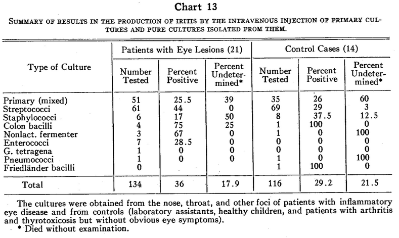
Of the total of 134 cultures from patients with eye disease, 36 percent produced iritis while 17.9 percent were undetermined. Of the total of 116 cultures from persons in the control group, 29.2 percent produced iritis in rabbits, while 21.5 percent were undetermined.
Toxicity, as measured by in-vitro tests, did not seem to be related to the iritis-producing power of streptococci and staphylococci. Seventy percent of the organisms which produced iritis gave positive toxicity reactions, whereas 60 percent of the strains which did not produce iritis also gave positive toxicity reactions.
It is concluded that, while iritis is produced in rabbit’s eyes by various cultures of bacteria, this property is not characteristic of any one bacterial genus, neither is it distinctly a property of cultures from patients with inflammatory eye diseases.
We wish to express our sincere appreciation to Dr. James M. Evans and Miss Adele Mayo for their cooperation in this study.
*The in-vitro toxicity tests referred to in this paper are listed in the following order: for staphylococci, hemolysis and coagulase tests18 and violet agar reaction;19 for streptococci, resistance to sodium bicarbonate and hexylresorcinol.21 In the staphylococcal reactions, toxicity is graded from negative to 4+. In the streptococcal tests, toxicity is graded from negative to 8+.
References Cited:
- Rosenow, E. C., and Nickel, A. C. “Elective localization in determining etiology of chronic uveitis.” Amer. Jour. Ophth., 1932, v. 15, p. 1.
- Maestro, T. “Oculotropismo sperimentale degli streptococchi.” Boll. d’ocul., 1935, v. 14, p. 1251.
- Zanettin, G. “Localizzazione elettiva dello stafilococco nell’occhio (Contributo sperimentale alla questione delle infezioni focali).” Ann. di ottal. e clin. ocul., 1933, v. 61, p. 20.
- —-“Infezioni focali e malattie oculari.” Ann. di ottal. e clin. ocul., 1934, v. 62, pp. 588, 695, and 786.
- Blanc, G., and Martin, L. A. “Iridocyclite expérimentale provoquée par virus typhique.” Compt. rend. Acad. d. sc., 1935, v. 200, p. 865.
- Cusumano, A. “Infezione focale e localizzazione secondaria nell’occhio (Contributo sperimentale sul tropismo elettivo batterico).” Rassegna Ital. d’Ottal., 1935, v. 4. p. 46.
- Wherry, W. B., and King, C. “Case illustrating local sensitization of eye to bacterial protein.” Jour. Med., 1927, v. 8, p. 85.
- de Andrade, L. “Experiments on the influence of injuries of the eye on localization of focal phenomena from tubercle bacilli introduced into the blood stream, and remarks on the question of sympathetic ophthalmia.” Klin. M. f. Augenh., 1934, v. 92, p. 350.
- von Herrenschwand, F. “Spirochaeten und Bacillus fusiformis bei akuter Konjunktivitis.” Zeit. f. Augenh., 1927, v. 62, p. 370.
- Brown, A. L. “Considerations underlying experimental production of uveitis.” Amer. Jour. Ophth., 1932, v. 15, p. 19.
- —-“Chronic uveitis. Bacteriologic and immunologic considerations.” Arch. of Ophth., 1934, v. 12, p. 730.
- Irons, E. E., Brown, E. V. L., and Nadler, W. H. “The localization of streptococci in the eye. A study of experimental iridocyclitis in rabbits.” Jour. Infect. Dis., 1916. v. 18, p. 315.
- Meisser, J. G., and Gardner, B. S. “Elective localization of bacteria isolated from infected teeth.” Jour. Amer. Dent. Assoc., 1922, v. 9, p. 578.
- Haden, R. L. “Elective localization in eye of bacteria from infected teeth.” Arch. Int. Med., 1923, v. 32, p. 828.
- Billings, F. Focal infection. New York, Appleton-Century Co., 1916.
- Berens, C., Connolly, P. T., and Chapman, G. H. “Focal infection in diseases of the eye. 1. Report of certain laboratory examinations.” Brit. Jour. Ophth., 1934, v. 18, p. 463.
- Rawls, W. B., and Chapman, G. H. “Experimental arthritis in rabbits. Comparison of the arthritis-producing ability of inagglutinable streptococci which resist the ‘bactericidal’ action of fresh, diluted, defibrinated guinea pig blood and those which are agglutinable but sensitive to the ‘bactericidal’ agent.” Jour. Lab. and Clin. Med., 1935, v. 21, p. 49.
- Chapman, G. H., Berens, C., Peters, A., and Curcio, L. “Coagulase and hemolysin tests as measures of the pathogenicity of staphylococci.” Jour. Bact., 1934, v. 28, p. 343.
- Chapman, G. H., and Berens, C. “Crystal violet agar as a differential medium for staphylococci.” Jour. Bact., 1935, v. 29, p. 437.
- Chapman, G. H., and Rawls, W. B. “Studies of streptococci I. Qualitative differences in resistance to various agents.” Jour. Bact., 1936, v. 31, p. 323.
- Chapman, G. H., and Curcio, L. “Studies of streptococci II. Quantitative differences in resistance to sodium bicarbonate and hexylresorcinol.” Jour. Bact., 1936, v. 31, p. 333.
- Brown, A. L. “Chronic uveitis. Bacteriologic and immunologic considerations.” Trans. Sec. Ophth. Amer. Med. Assoc., 1934, p. 111.
- Alagna, G., and Tallo, F. “Sulla diagnosi dei foci tonsillari e sul tropismo elettivo dei batteri in essi contenuti; Contributo clinico-sperimentale.” Arch. Ital. di Otol. Rinol. e Laringol., 1935, v. 47, p. 112.
- Collins, E. T., and Mayou, M. S. Pathology and bacteriology of the eye. Ed. 2, Philadelphia, P. Blakiston’s Son and Co., 1925, p. 557.
- Meller, J. “Nachweis von Tuberkelbazillen bei Uveitis durch Kultur aus dem Gewebe des Augeninnern.” Zeit. f. Augenh., 1932, v. 77, p. 1.