
Access to all articles, new health classes, discounts in our store, and more!
Localization in Animals of Streptococci From Cases of Epidemic Hiccup, Encephalitis, Spasmodic Torticollis and Chorea
Read before the Section on Nervous and Mental Diseases at the Seventy-Eighth Annual Session of the American Medical Association, Washington, D. C., May 19, 1927. Published in the Archives of Neurology and Psychiatry, Vol. 19, March 1928, pp. 424-436.
* * *
In a series of reports published in the medical literature during the last eleven years, I have emphasized1 the importance in the etiology of diseases of the nervous system of certain streptococci having peculiar localizing power. The experimental results, while not always conclusive proof of etiologic relationship in each of the diseases studied, have furnished a good working hypothesis which already has led to a better understanding of the cause and management of the diseases studied.
In 1916, I reported2 a series of experiments in which lesions in the spinal cord or brain of animals, characteristic in situation, were produced by the intravenous injection of cultures containing streptococci from atria of the infection in cases of multiple sclerosis and transverse myelitis. Similar experiments in cases of neuralgia and multiple neuritis produced instead lesions chiefly in the dorsal nerve roots and peripheral nerves, respectively, in a high percentage of animals inoculated. These experiments suggested strongly that specific or elective localizing power of streptococci, inherent or acquired, must be an important factor in the production of various diseases of the nervous system, just as I had shown this to be the case in other diseases. The work led to the isolation of the pleomorphic streptococcus in epidemic poliomyelitis in the same year and, in 1921, to a study of the etiology of epidemic encephalitis.
In encephalitis it was found that intracerebral injection oi small amounts of suspensions (in sodium chloride solution) of material expressed from tonsils, swabbed from the nasopharynx or aspirated from pyorrhea pockets, and of the corresponding primary cultures in broth, was more likely to produce characteristic symptoms than intravenous injection, and often served to separate the causative streptococcus from contaminating organisms and other streptococci which did not possess neurotropic power. By this method, the condition of profound lethargy never before seen or described in rabbits, together with other concomitant symptoms manifested by the patient, was produced with a streptococcus from a fatal case of epidemic encephalitis. In this case, the streptococcus was isolated from infected teeth, from the filtrate of the nasopharyngeal washings, from the blood during life, from an emulsion of the base of the brain after death and from a filtrate of the emulsion of the brain. lt was demonstrated in the lesions of the brain and proved to be absent in the normal brain tissue. It was isolated from the brain and demonstrated in the lesions of positive animals. Similar experiments with material from normal persons failed to yield this organism. Since that time, similar results have been reported with culturally and immunologically identical streptococci in eighty-one cases of various forms of encephalitis. An agglutinating and curative serum has been prepared and used to identify suspected strains and in the treatment of a series of patients with encephalitis. The streptococcus isolated in these cases often varied greatly in size, depending on the medium in which it was cultivated. Exceedingly small forms were found, especially in old cultures in Noguchi’s medium, and large, ordinary-sized, and exceedingly small forms were demonstrated in the lesions in a series of fatal cases of encephalitis by Rosenow and Jackson.3
Moreover, epidemic hiccup, that strange condition manifested by changes in character and posture, associated often with extreme respiratory arrhythmia that may follow attacks of encephalitis, spasmodic torticollis and chorea, has been reproduced, in the main, in animals with similar streptococci isolated from various atria of infection in these diseases. The experiments with epidemic hiccups were especially illuminating. Spasms of the diaphragm were produced with great regularity, in rabbits, guinea-pigs, and monkeys (a condition not known to occur spontaneously in these animals) with cultures from the nasopharynx or tonsils in cases occurring in a series of outbreaks. It was found that spasms of the diaphragm could be produced not only after injection of the living streptococcus, but also when the organism was killed by heat or formaldehyde, and when corresponding broth-culture filtrates were used after the living cultures had lost this power, the dead bacteria and filtrate also failed to cause diaphragmatic spasms. Likewise, filtrates of nasopharyngeal washings from patients at the time of the attack caused spasms of the diaphragm and of other muscles, while filtrates obtained after recovery and after the specific streptococcus had disappeared, were without effect.
Another striking example of the extreme specificity of the streptococcus and the variation in this property under certain conditions was found in experiments performed in a series of cases presenting varying degrees of neuromyelo-encephalitis which occurred during and after an epidemic of hiccup. Localizations, with lesions strikingly like those of the patient in [this] situation, occurred in animals injected with material from the nasopharynx or tonsils. These results suggested strongly that the changing character in the epidemic was due to changes in the isolated streptococcus, since the symptoms and lesions in the experimental animal simulated so closely those of the patient in different stages of the epidemic.
A number of circumstances have retarded the general acceptance of the significance of these studies, especially in encephalitis, and reports on further studies are greatly needed. The work stood unconfirmed until recently, when Evans and Freeman4 reported observations in several cases of epidemic encephalitis which in important respects paralleled those which I have reported. This is true especially as regards the site of isolation of the streptococcus, its sensitiveness to oxygen, its pleomorphism, filtrability and its effect in rabbits. The streptococcus isolated in these diseases morphologically and culturally resembles other streptococci which have only slight if any neurotropic properties. Hence, extraordinary proof of causal relationship is required. The streptococcus tends to lose specific localizing power on artificial cultivation as well as on successive passage through animals, and reproduction of the disease is thus rendered difficult. The streptococcus is highly sensitive to oxygen and may easily be missed. Emulsions of the brain in fatal cases may not contain a sufficient number of living organisms to affect animals, for it has been shown that if cultures or nasopharyngeal washings are too highly diluted, symptoms do not develop; this also has been well shown in the recent work of Evans and Freeman. The production of antibody is slight or not demonstrable, especially in afebrile cases, and hence agglutination or other serum reactions with suspected organisms are generally negative. Varying numbers of rabbits, the species of choice in these experiments, have been found to have spontaneous lesions of the brain resembling encephalitis. This objection, however, has been met in my own studies, because sections of the brain of numerous control animals showed the incidence to be very low (much lower than in the animals into which injections were made); the relation of the injected organism to the lesions was demonstrated microscopically, and similar lesions were produced in guinea-pigs, mice and monkeys, in which these lesions do not occur spontaneously. Moreover, the spontaneous disease is symptomless while, in my experiments, symptoms were pronounced and often simulated those in the patient from whom the strain was obtained. Because certain workers have succeeded in producing typical lesions in rabbits with virus and have isolated a small anaerobic organism resembling the globoid bodies of poliomyelitis, it is thought by many that streptococci cannot have etiologic significance. This contention, however, is no longer tenable, for Evans has recently reported the successful isolation of bacteria, including the streptococcus having neurotropic power, from the so-called viruses of encephalitis and herpes.
I shall now report further results on the localizing and symptom-producing power of the streptococci isolated in several diseases of the nervous system, and record the effects in animals of filtrates of broth cultures and nasopharyngeal washings.
The method of study was similar in the different diseases studied, and has been described in detail previously. It suffices to state here that the results summarized in tables 1 and 2 were obtained chiefly by injecting far forward into the right frontal lobe, from two to six rabbits, 0.1 cc. and 0.2 cc. of nasopharyngeal swabbings, or of pus expressed from tonsils suspended in 2 cc. of gelatin (0.1 per cent) Locke solution or these amounts of the primary cultures in dextrose brain broth, or of cultures from the apexes of extracted, devitalized teeth, diluted 1:10. Ether anesthesia was used in these experiments. Blood-agar platings were always made of the material injected to determine the viability and the kind of bacteria injected. The animals were observed as a routine twice daily and more often if active symptoms developed. In many instances, material from patients having a different disease was injected on the same day into animals derived from a common supply; this served as a check on susceptibility of different breeds or lots. Exact dosage in this work according to weight or age, as in intracerebral injection of the virus of poliomyelitis in monkeys, is not essential.
Table 1.—Mortality and Percentage Incidence of Symptoms in Animals Following Intracerebral Inoculation
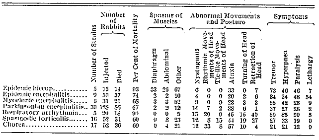
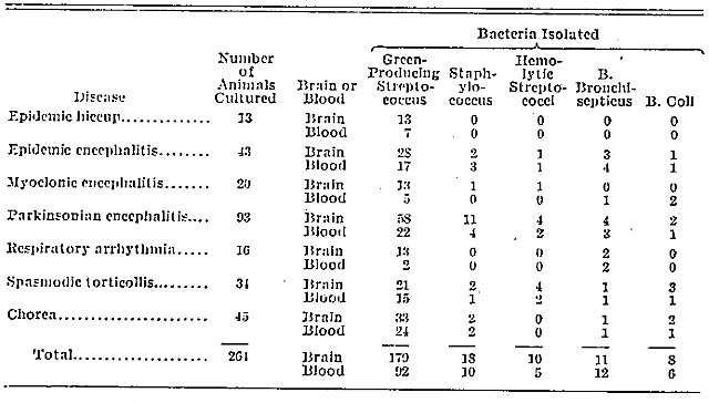
Table 2.—Bacteria Isolated from Brain and Blood of Inoculated Animals
Clinical Results
There was no reasonable doubt as to the correctness of the diagnosis in the cases studied. Nearly all of the patients were seen in consultation with members of the staff of the Mayo Clinic and were considered as suitable for the possible isolation of the causative organism through intracerebral inoculation of animals with material from tonsils, throat or infected teeth. Definite evidence of focal infection was found in most of the cases, and in some this evidence was marked, especially in the teeth. All of the cases in which inoculation of the animals was performed are included in tables 1 and 2, irrespective of whether the results were positive or negative.
In certain instances, the results in animals were used as evidence for or against the removal of tonsils or other foci of infection, although in most cases removal was advised irrespective of the laboratory results. Autogenous vaccines were prepared, in many cases, from the streptococcus isolated from the brain of animals with positive reactions. The clinical results from the removal of foci of infection and from the use of the vaccine in these and other cases will be reported later. Relief was obtained frequently in cases in which the symptoms had persisted for months, while in others a progressive course was arrested, and in still others apparent benefit was not evident.
The age of the patients ranged from 5 to 68 years in the different groups and was not of particular significance. The duration of symptoms in the cases of persistent hiccup ranged from two days to twenty-one months; in the group with epidemic encephalitis, from twelve days to five months; in that with myoclonus, from ten weeks to nine months; in that with the parkinsonian encephalitis, from one week to ten years; in that with respiratory arrhythmia, from six weeks to two years; in that with spasmodic torticollis, from six months to ten years, and in that with chorea, from two weeks to four months.
Results of Experiments on Animals
The number of cases studied in the different groups varied from 5 to 30, the number of animals injected from 15 to 128, and the mortality rate from 60 to 93 per cent (table 1). The incidence of the different symptoms is recorded in percentages, hence the figures are directly comparable. In order to simplify the table, the fractions below 0.5 per cent are dropped, while the next higher figure is used if 0.5 or above. Certain symptoms, such as ataxia, tremors, hyperpnea and paralysis are relatively common in most of the diseases studied, especially in fatal cases, and, correspondingly, the general incidence of these symptoms is high in the experimental animals. Certain other symptoms, such as spasms of the diaphragm and abdominal muscles, lethargy, rhythmic and ticlike movements and rotation of the head, occur more rarely in patients affected with these diseases, and, in agreement with this fact, their general incidence is low in the animals that have received injections 2 (table 1).
The incidence in the animals of the symptoms most characteristic of the different groups was often strikingly high, and in some cases simulated the condition in the patient so closely as to make one feel certain that the material injected must have contained the etiologic agent. This occurred especially in cases in which the patients’ symptoms were of recent origin or in which exacerbations of symptoms of long duration had occurred shortly before the study was made. Spasms of the diaphragm were noted in 33 per cent of all the animals injected with material from patients with persistent hiccup, which was in sharp contrast to the next highest incidence (3 per cent) of the symptoms in the other groups. Spasms of the muscles (67 per cent) was highest in the groups with persistent hiccup and in that with myoclonus (52 per cent); it was also relatively high in the other two groups in which this was an outstanding symptom, namely, in spasmodic torticollis (23 per cent) and in chorea (21 per cent). In the cases of myoclonic encephalitis the animals sometimes manifested spasms of the same group of muscles as was affected in the patient from whom the material was obtained (figs. 1 and 2).
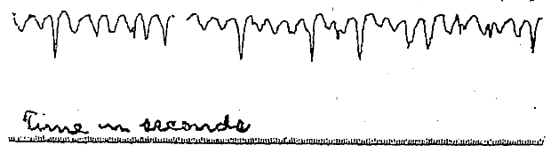
Fig. 1.—Curve showing spasms of the abdominal muscles in a case of myoclonic encephalitis of three months’ duration. Note the spasms occurring irregularly at the end of inspiration.
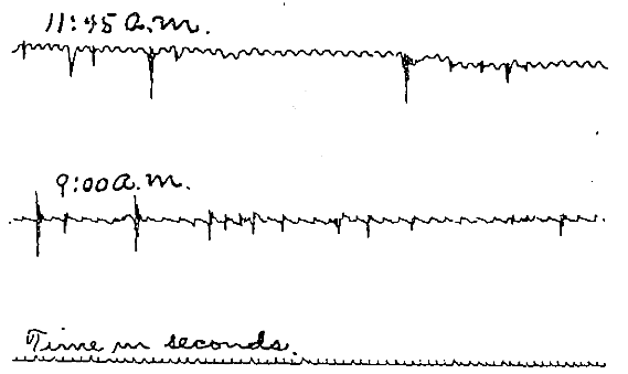
Fig. 2.—Curve showing spasms of the abdominal muscles in a rabbit injected intracerebrally, the day previously, with 0.2 cc. of a suspension from the nasopharynx in the case referred to in figure 1.
As a prominent symptom, lethargy was noted in 54 per cent of the animals injected with material from patients with epidemic encephalitis; this was in striking contrast to the next highest figure of 9 percent in the groups in which this symptom was not encountered. It varied in intensity and duration from slight somnolence lasting for a day or two to deep sleep continuing for a number of days, from which the animal could be aroused only with difficulty. In one case of epidemic encephalitis, typical symptoms developed in all of four rabbits injected with material obtained directly from the tonsils and the nasopharynx and in all of three injected with streptococcus isolated from the brain of one of these. Moreover, the fresh filtrate of nasopharyngeal washings which yielded the characteristic streptococcus in cultures in dextrose-brain broth and meat-mash medium produced lethargy in both of the two rabbits injected. This symptom developed within several; hours after injection, indicating the presence of a toxin having specific localizing or symptom-producing power. Cultures from the brain of these animals yielded the streptococcus, and emulsions injected directly into three other rabbits produced lethargic symptoms in two and encephalitis in all.
On the basis of these striking results, three additional rabbits were injected with the filtrate after it had been kept for four days in the ice box. All the animals remained well, and the cultures proved sterile.
The results in two additional cases of epidemic encephalitis studied at the same time were similar to those cited in the preceding case. Injections of suspensions of the pus from the tonsils of both patients produced lethargic symptoms of encephalitis in animals. The filtrate from a mixture of the suspension of the pus expressed from the tonsils, which proved sterile in culture, produced pronounced symptoms of lethargy in both of two rabbits given intracerebral injections. This began one hour after injection, continued for several days in both animals, and then disappeared.
In the group with parkinsonian encephalitis a particularly high figure was not noted among the symptoms listed in table 1, although ataxia (28 per cent) and tremors (37 per cent) were often pronounced. Slowness of movement, increased muscular tonus, rigidity in various regions and drooling of saliva, especially in animals that lived for some time, were not infrequently noted. The results in one case in this group, that of a patient with bulbar paralysis, manifested by weakness of the muscles of the tongue, throat and neck, were especially interesting. Two rabbits injected with the primary culture from an infected tooth of the patient and two with the emulsion of the brain of one of the rabbits injected with the primary culture manifested a bulbar type of paralysis beginning with weakness of the muscles of the neck. Three of four rabbits given injections with suspensions of the pus from the tonsils on two occasions, four days apart, also developed encephalitis manifested especially by weakness of the muscles of the neck, ears and fore extremities.
Hyperpnea and irregularity in breathing, often extreme, and sometimes associated with an expiratory grunt, were noted in 85 per cent and retraction of the head in 40 per cent of the animals inoculated with material from patients manifesting respiratory arrhythmia; this corresponded to the most striking symptoms noted in this group of patients. The results in one of these cases, that of a patient with extreme respiratory arrhythmia and hyperpnea, deserve special emphasis. Five of six rabbits injected with the undiluted and diluted (1:100) suspension of nasopharyngeal swabbings manifested pronounced respiratory symptoms and died, seemingly of bulbar paralysis. Cultures from the brain yielded the streptococcus in all. The strain from one of these rabbits was subcultivated rapidly three times and then grown in hormone broth for seven days. This exhaust culture was then filtered and the sterile filtrate injected intracerebrally into two rabbits, one receiving 2 cc., the other 1.5. The respirations became greatly increased in both, several hours after injection, but disappeared over night. Reinjection the following day produced hyperpnea as the chief symptom. Three days after the first injection, 7 cc. of the filtrate was injected intravenously in each of the rabbits; hyperpnea again appeared, lasted for several hours and then disappeared.
In sixteen cases in the spasmodic torticollis group, the symptoms induced were often strikingly like those characteristic of this condition in patients. Spasms of the muscles were noted in 23 per cent of the animals injected, ataxia, often extreme, in 44 per cent, tic-like movements of the head in 15 per cent, retraction of the head in 27 per cent, and turning of the head, due to tonic contraction of the cervical muscles, lasting often for weeks or months, in 19 per cent. The results with cultures from one of these cases of marked torticollis of six months’ duration were most interesting. Symptoms developed in five of seven rabbits injected with suspensions of nasopharyngeal swabbings in dilutions as high as 1:1,000 and in two of three rabbits injected with material from infected tonsils. A filtrate from nasopharyngeal washings produced similar effects in two of three rabbits given injections, and heat-killed primary culture in seven of eight rabbits. Lethargy and hyperpnea were not observed. The similarity of patients’ symptoms to those of the animals given injections was marked (fig. 3).
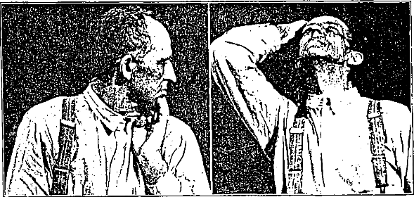
Fig. 3.—Turning and retraction of the head and attempts at their control in a patient with marked spasmodic torticollis of six months’ duration.
In the chorea group, ataxia, often extreme, was noted in 57 per cent of the animals given injections, rhythmic movements of the head in 33 per cent and spasms of the muscles in 21 per cent; these symptoms were in striking agreement with those commonly present in patients suffering from this disease.
Control inoculations with suspensions and culture of streptococci without virulence or neurotropic power, sterile filtrates, sodium chloride solution, and broth, have been made in a large number of animals, always without apparent effect.
The incidence of the isolation of different bacteria from the brain and blood of the animals injected with material from patients in the different groups, and in which cultures were made after death, is given in table 2. Most of these died from the effect of the injection, although some were chloroformed for examination. The total incidence of the isolation of the green-producing streptococcus is much higher in each of the groups than of the staphylococcus, hemolytic streptococcus, Bacillus bronchisepticus, and Bacillus coli. Cultures from the blood yielded the streptococcus approximately only in one third of the animals from which cultures were taken, and hence the streptococcus was of low general virulence. Blood-agar plates usually showed a relatively small number of colonies of this organism and often no growth when inoculations in dextrose-brain broth yielded pure cultures. The number of colonies of the streptococcus from the brain on blood-agar plates was usually very large in the case of the animals that died soon after inoculation and very small in the animals that lived for a long time or that were anesthetized as they were recovering. Cultures from the brain were also frequently negative on blood-agar platings, when positive in dextrose-brain broth.
The green-producing streptococci isolated from the positive animals in the different diseases studied are very much alike morphologically and in cultural features, virulence and immunologic properties. The chief difference between the strains lies in their specific effects in animals on isolation, a property quickly lost, especially on aerobic cultivation.
The observations at necropsy were often largely negative and not characteristic of any of the diseases studied. Lesions at the point of injection were usually absent, although occasionally localized softening, with or without hemorrhage, was found. Suppurative meningitis almost never occurred, and when it did, it was nearly always due to hemolytic streptococci. Moderate or slight clouding of the cerebrospinal fluid, clouding of the pia and congestion of the vessels of the meninges were found regularly in the animals that succumbed soon after injection but were rare in the animals that lived for a longer time. Gross hemorrhages into the pia or substance of the brain and spinal cord were rare and occurred most commonly in the medulla or pons following injection of material from patients manifesting bulbar symptoms. Localized clouding and edema of the pia over the anterior aspect of the medulla, pons and cervical cord occurred especially in the animals injected with material from patients suffering from epidemic hiccup, spasmodic torticollis and respiratory arrhythmia. Besides edema of the lungs and infrequent dilatation and postmortem digestion of the stomach, lesions of the viscera were rare following intracerebral inoculation of material from different groups of cases. Lesions of the heart valves or endocardium, however, were commonly noted following intracerebral inoculation of the streptococcus from foci of infection or from the throats of patients suffering from chorea. This condition practically never occurred following inoculation of the streptococcus in cases of encephalitis and allied conditions. The microscopic changes produced were almost similar in the different groups and were characteristic in some of the diseases studied only as far as the site was concerned. Cellular infiltration varied greatly according to the duration of the experiment, size of dosage and virulence of the strain injected. Leukocytic and round cell infiltration of the pia was most marked surrounding the blood vessels in the sulci. Focal hemorrhages in the brain and medulla, often perivascular, associated with a variable degree of leukocytic and round cell infiltration, and similar areas of infiltration unassociated with hemorrhage, were usual observations in the animals that succumbed or were anesthetized during the first five days after injection. The streptococcus was readily demonstrated in these lesions. As the duration of the experiment became longer, hemorrhages were less common, the leukocytes were displaced by round cells, especially in the perivascular spaces in the midbrain (Fig. 4), and the diplococci became fewer and more irregular in size (large and small forms often occurring side by side and sometimes in the same chain); they usually occurred in groups in perivascular spaces (Fig. 5). The proportion of leukocytes and round cells varied greatly according to the site of the lesion; the former often predominating in the pia and the latter in the perivascular spaces in the depths of the brain substance.
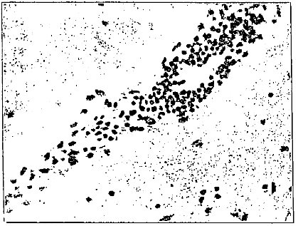
Fig. 4.—Typical perivascular, round cell infiltration in the midbrain of a rabbit injected, eleven days previously, with a suspension of the nasopharyngeal swabbings from a patient with typical lethargic encephalitis; X 250.
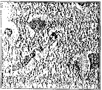
Fig. 5.—Streptococci in a perivascular space adjacent to the area of infiltration shown in figure 4; X 1.000.
Owing to the widespread presence of microscopic changes, or for unknown reasons, it was not always possible to correlate accurately the lesions found after death with particular symptoms noted during life.
Comment and Conclusions
Another series of experiments with animals performed with material from foci of infection or throats of patients suffering from various diseases of the nervous system is reported. The results corroborate and extend the former observations. The streptococci isolated in each of these conditions, while much alike, possess very different localizing and symptom-producing power when injected intracerebrally into animals and readily lose this property, especially on aerobic cultivation. The specific localizing power of the streptococcus, it would seem, is dependent, in part at least, on the production of a toxic or poison that has specific effects. Lethargy, hyperpnea with marked arrhythmia, and the symptoms characteristic of spasmodic torticollis have been produced with filtrates from nasopharyngeal washings, filtrates of suspensions of pus from tonsils and filtrates of old cultures of the specific streptococcus in each of these conditions. The fact that the streptococcus, having specific effects and the power to produce a specific poison, has been isolated long after onset indicates that the late manifestations of encephalitis and allied conditions are not sequels but are due to the activity of the causative organisms or its toxin, as has been especially emphasized by Freeman.
It is of course realized that the results obtained are not absolute proof of the etiologic relationship of the streptococcus isolated. None of the patients in this group died, and hence search for the organism in lesions could not be made. Agglutination experiments, especially with the serum of patients and the suspension of the suspected organism, while sometimes positive in low dilutions, were not so regular enough to be convincing. The benefits which follow the removal of foci proved to contain the organism and the use of autogenous vaccines, while often striking, do not occur regularly enough to prove etiologic relationship.
In consideration of all the facts, especially the extremely specific effects obtained in some cases with the streptococcus isolated, and its specific toxin or poison-producing power reported herewith, the tentative conclusion seems warranted that this organism has etiologic significance in each of the diseases studied.
References Cited:
- Fielmholz, H. F.. and Rosenow, E. C.: Three Cases of Acute Encephalitis Treated with Specific Serum, J. A. M. A. 79:2068 (Dec. 16) 1922. Rosenow, E. C.: Elective Localization of Streptococci, J. A. M. A. 65:1687 (Nov. 13) 1915; Experimental Studies on the Etiology of Encephalitis; Report of Findings in One Case, ibid. 79:443 (Aug. 5) 1922; Specific Serum Treatment of Epidemic (lethargic) Encephalitis; Further Results, ibid. 80:1583 (June 2) 1923; Experimental Observations on the Etiology of Chorea, Am. J. Dis. Child. 26:223 (Sept.) 1923; The Production of Spasms of the Diaphragm in Animals by Living Cultures, Filtrates, and the Dead Streptococcus from Cases of Epidemic Hiccup, J. Infect. Dis. 32:72 (Jan.) 1923; Streptococci in Relation to the Etiology of Epidemic Encephalitis: Experimental Results in Eighty-One Cases, ibid. 34:329 (April) 1924; Experiments on the Etiology of Respiratory Arrhythmias Following Epidemic Encephalitis, Arch. Neurol. & Psychiat. 11:155 (Feb.) 1924; Experimental Studies Indicating an Infectious Etiology of Spasmodic Torticollis, J. Nerv. & Jent. Dis. 59:1, 1924; Further Studies on the Etiology of Epidemic Hiccup (Singultus and Its Relation to Encephalitis, Arch. Neurol. & Psychiat. 15:712, 1926; Neuromyelo-Encephalitis During and Following an Epidemic of Hiccup; Diverse Localization of Streptococci, ibid. 16:21 (July 26); Diaphragmatic Spasms in Animals Produced with a Streptocococcus from Epidemic Hiccup; Preliminary Report, J. A. M. A. 76:1745 (Jne 18) 1921. Rosenow, E. C., and Wheeler, G. W.: The Etiology of Epidemic Poliomyelitis, J. Infect. Dis. 22:281 (April) 1918.
- Rosenow, E. C.: Elective Localization of Bacteria in Diseases of the Nervous System, J. A. M. A. 67:662 (Aug. 26) 1916.
- Rosenow, E. C., and Jackson, G. II.: Microscopic Demonstration of Bacteria in the Lesions of Epidemic Lethargic Encephalitis, J. Infect. Dis. 32: 144 (Feb.) 1923.
- Freeman, Walter: Chronic Epidemic Encephalitis, J. A. M. A. 87:1601 (No. 13) 1926. Evans, Alice C., and Freeman, Walter: Studies on the Etiology of Epidemic Encephalitis: I. The Streptococcus, U. S. Public Health Rep. 41: 1065, 1926. Evans, Alice C.: Studies on the Etiology of Epidemic Encephalitis: II. Virulent Bacteria Cultivated from So-Called Herpetic and Encephalitis Viruses. U. S. Public Health Rep. 42: 171, 1927.